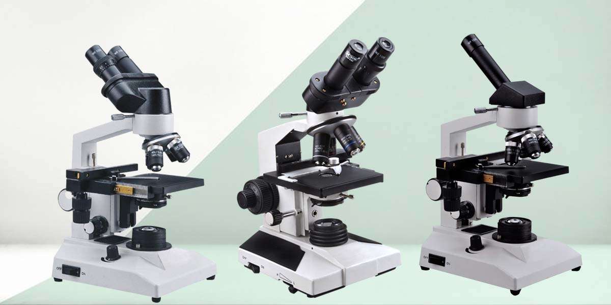
A laboratory microscope is a precision optical instrument designed to magnify and resolve fine details of specimens that are not visible to the naked eye. It is a fundamental tool in various scientific fields such as biology, medicine, materials science, and chemistry. Here’s a detailed overview of its components, types, working principles, and uses:
Key Components of a Laboratory Microscope
Eyepiece (Ocular Lens)
- Located at the top of the microscope.
- Usually provides 10x magnification.
Objective Lenses
- Mounted on a rotating nosepiece.
- Provide varying levels of magnification (commonly 4x, 10x, 40x, 100x).
- The total magnification is the product of the eyepiece and objective lens.
Stage
- A flat platform where the slide is placed.
- Includes clips or a mechanical holder to secure the slide.
Light Source (Illuminator)
- Positioned beneath the stage.
- Provides light to illuminate the specimen.
Condenser Lens and Diaphragm
- Focuses and controls the amount of light reaching the specimen.
Coarse and Fine Focus Knobs
- Used to bring the specimen into sharp focus.
- Coarse adjustment moves the stage quickly, while fine adjustment allows for detailed focusing.
Arm and Base
- Provide support and stability for the microscope.
Types of Laboratory Microscopes
Light Microscope (Optical Microscope)
- Uses visible light and lenses to magnify specimens.
- Common in schools and basic labs.
- Subtypes:
Compound Microscope: High magnification, uses multiple lenses.
Stereo Microscope (Dissecting Microscope): Low magnification, for viewing surface details.
Electron Microscope
- Uses beams of electrons instead of light.
- Much higher magnification and resolution.
- Subtypes:
Transmission Electron Microscope (TEM): Views internal structures.
Scanning Electron Microscope (SEM): Views surface structures in 3D.
Fluorescence Microscope
- Uses high-intensity light to excite fluorescent dyes in the sample.
- Useful in cell biology and medical diagnostics.
Working Principle
- Illumination: The light source illuminates the specimen on the stage.
- Magnification: Light passes through the objective and eyepiece lenses, magnifying the image.
- Focusing: Coarse and fine knobs adjust the distance between the lenses and the slide for a clear image.
- Viewing: The magnified image is viewed through the eyepiece or projected onto a screen or camera.
Applications of Laboratory Microscopes
- Biology: Observing cell structures, microorganisms, and tissues.
- Medicine: Diagnosing diseases through blood, tissue, and urine analysis.
- Materials Science: Analyzing the microstructure of metals, polymers, and composites.
- Forensics: Examining hair, fibers, and other microscopic evidence.
- Education: Teaching students about cells, microorganisms, and histology.
Benefits of a Laboratory Microscope
Enhanced Visualization of Microscopic Structures
- Main Benefit: Microscopes magnify objects too small to be seen with the naked eye.
- Impact: Scientists and researchers can observe individual cells, bacteria, viruses, and organelles.
- Example: A biologist can study the nucleus, mitochondria, and other organelles inside a cell.
Advancement in Medical Diagnostics
- Main Benefit: Enables detailed examination of blood, tissues, urine, and other biological samples.
- Impact: Helps detect diseases such as cancer, infections, and genetic disorders at an early stage.
- Example: Pathologists use microscopes to identify abnormal cells in a biopsy to diagnose cancer.
Supports Scientific Research and Discovery
- Main Benefit: Fundamental tool for research in biology, chemistry, physics, and material science.
- Impact: Leads to breakthroughs in genetics, microbiology, pharmacology, and nanotechnology.
- Example: Microscopes were crucial in the discovery of DNA structure and bacterial behavior.
Improves Science Education
- Main Benefit: Visual learning tool in schools and universities.
- Impact: Helps students understand biological structures and processes in real-time.
- Example: Students learn to identify plant and animal cells, encouraging hands-on learning.
Quality Control and Manufacturing Precision
- Main Benefit: Used in industrial labs for quality inspection of materials and components.
- Impact: Ensures the accuracy and integrity of manufactured products, especially in electronics and pharmaceuticals.
- Example: Microscopes are used to inspect circuit boards for microscopic defects.
Aids in Genetic and Cellular Research
- Main Benefit: Allows observation of cell division, mutation, and interactions at the cellular level.
- Impact: Facilitates genetic engineering, stem cell research, and regenerative medicine.
- Example: Researchers use fluorescence microscopes to track specific proteins within cells.
Exploration of Microorganisms
- Main Benefit: Critical for studying bacteria, fungi, viruses, and protozoa.
- Impact: Contributes to disease control, vaccine development, and food safety.
- Example: Microbiologists identify pathogens in water samples to prevent contamination.
Forensic and Crime Investigations
- Main Benefit: Examines trace evidence like hair, fibers, and biological fluids.
- Impact: Helps solve crimes by linking suspects to crime scenes.
- Example: Forensic scientists use microscopes to match hair samples or analyze gunshot residues.
Drives Innovation in Nanotechnology
- Main Benefit: Enables observation and manipulation at the nanometer scale.
- Impact: Essential in developing nanomaterials, drug delivery systems, and molecular machines.
- Example: Scanning electron microscopes are used to design new drug carriers at the nanoscale.
Environmental Monitoring and Research
- Main Benefit: Used to analyze soil, water, and air samples for pollutants and microbes.
- Impact: Supports conservation efforts and environmental health.
- Example: Scientists examine plankton and microorganisms in water to assess ecosystem health.
Role of Laboratory Microscope in the Health Industry
Medical Diagnosis
Primary Role:
- Microscopes allow healthcare professionals to examine cells, tissues, and bodily fluids to identify diseases at the microscopic level.
Examples:
- Blood Smear: Detects anemia, malaria, leukemia.
- Urine Analysis: Identifies infections, crystals, or kidney disease.
- Histopathology: Examines biopsies for signs of cancer, inflammation, or other abnormalities.
Impact:
- Early and accurate diagnosis of diseases.
- Determines the severity and stage of conditions like cancer.
Microbiology and Infection Control
Primary Role:
- Identifies microorganisms like bacteria, viruses, fungi, and parasites that cause disease.
Examples:
- Staining and viewing bacteria in sputum, blood, or wound samples.
- Tracking antibiotic resistance through bacterial morphology.
Impact:
- Guides the correct use of antibiotics.
- Helps control outbreaks of infectious diseases.
Pathology and Histopathology
Primary Role:
- Examines tissue samples (biopsies) to detect abnormal cell growth, inflammation, and degeneration.
Examples:
- Cancer detection through tumor biopsies.
- Liver and kidney tissue evaluation for chronic diseases.
Impact:
- Critical in oncology, determining cancer type and grade.
- Guides surgical decisions and treatment plans.
Medical and Allied Health Education
Primary Role:
- Teaches students to recognize normal and abnormal microscopic structures in human tissues and fluids.
Examples:
- Viewing red and white blood cells.
- Identifying epithelial tissue or bone cells.
Impact:
- Builds diagnostic skills in future doctors, nurses, and lab technicians.
- Strengthens understanding of human anatomy and disease processes.

